
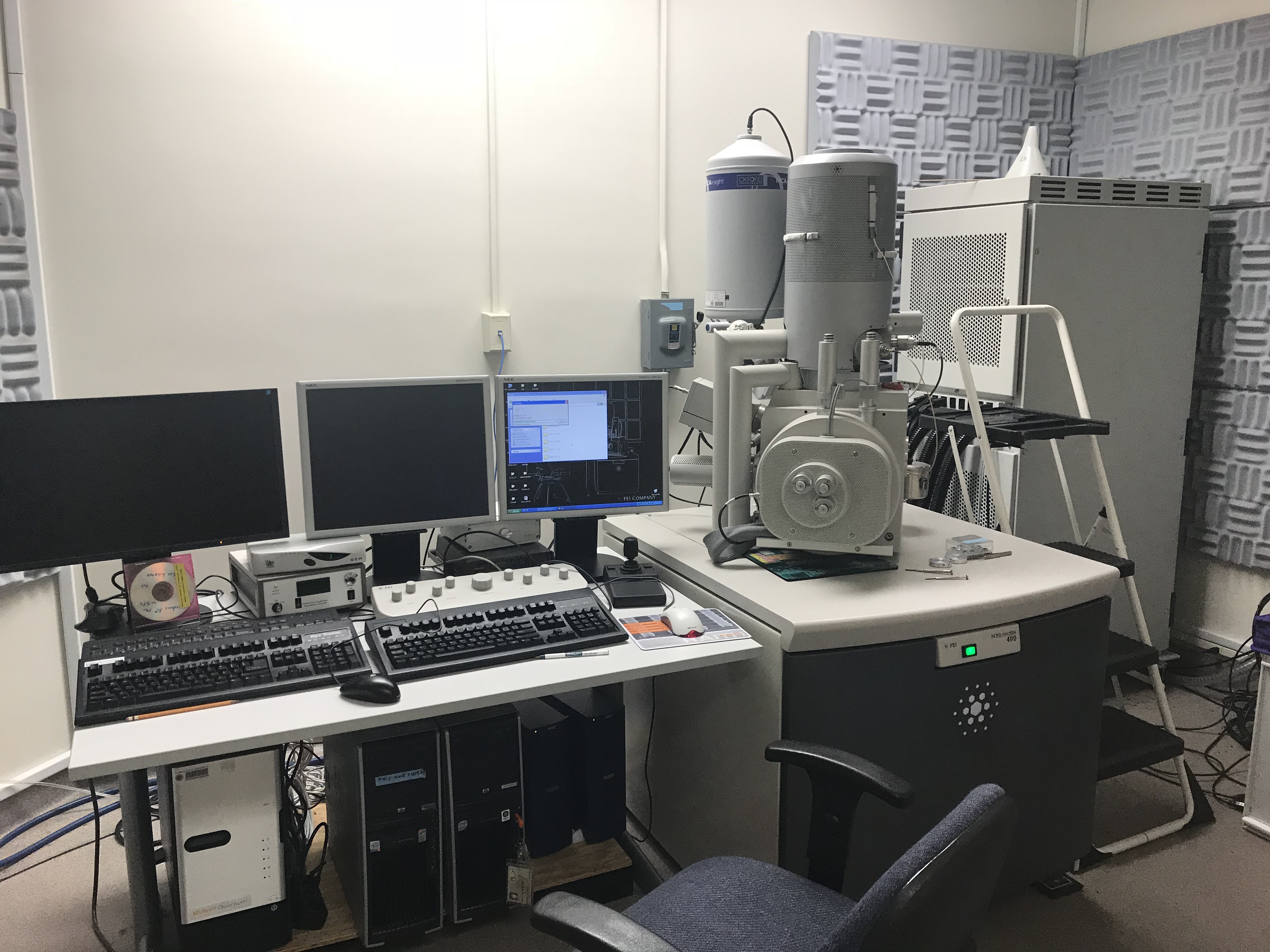
Bio Unit 1: Room 119C
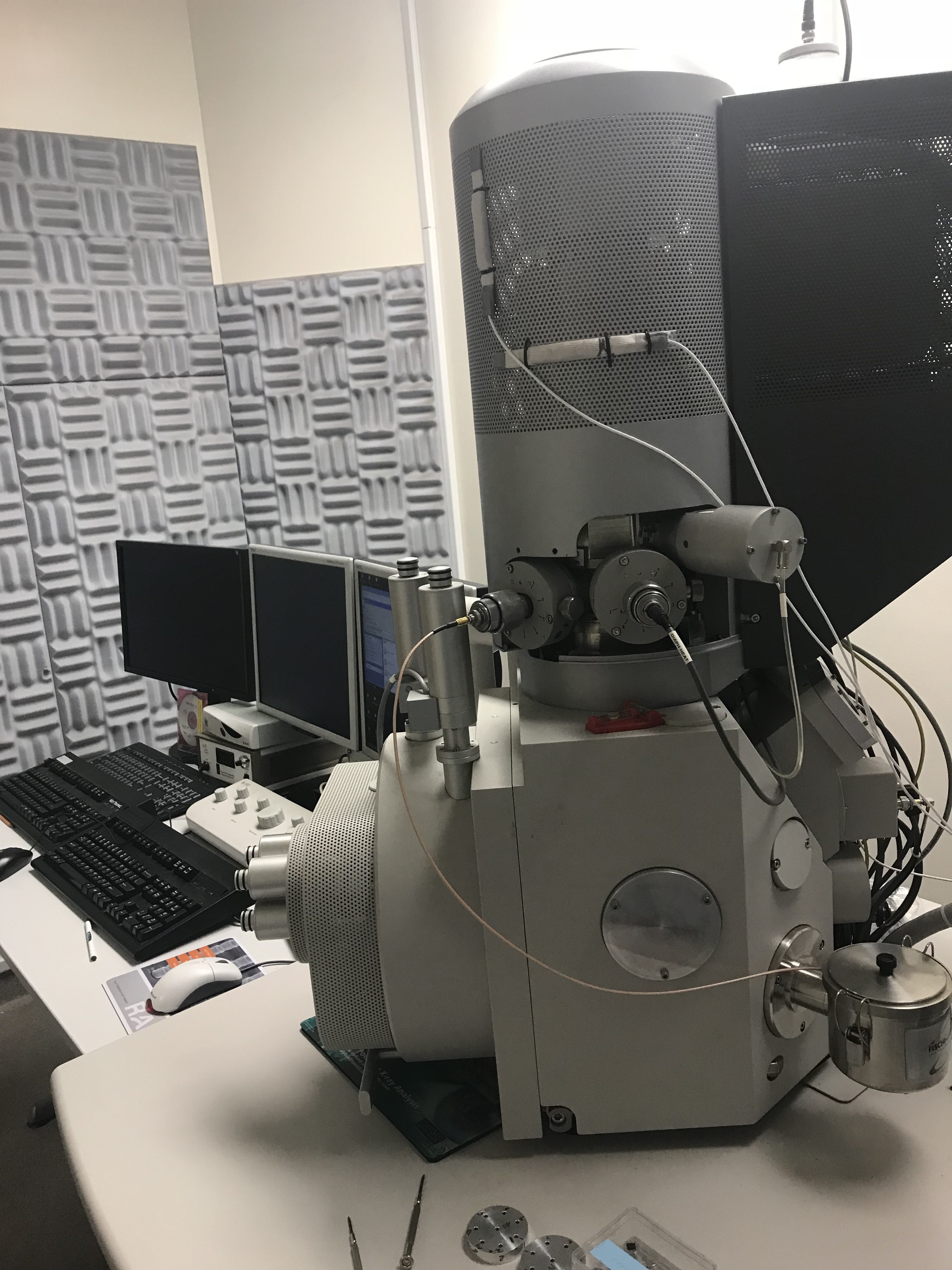
The Nova NanoSEM is an ultra-high resolution Low Vacuum Schottky Field Emission Scanning Electron Microscope (FEG-SEM). The instrument combines a field emission electron source, oil-free vacuum system, magnetic immersion final lens, heated objective aperture.
Specifications:
- 1.5 nm at 10 kV (Helix Detector)
- 1.8 nm at 3 kV (Helix Detector)
- In-lens SE (TLD-SE)
- In-lens BSE (TLD-BSE)
- Everhardt-Thornley SED
- Low vacuum SED (LVD)
- UHR low vacuum SED (Helix Detector)
- Low vacuum TV-rate solid-state BSED (GAD)
- Solid-state BSED for high vacuum
- STEM Detector
- IR-CCD camera for in-chamber viewing
- X = 100mm
- Y = 100mm
- Z = 60mm
- T = -5 degrees to +70 degrees
- R = 360 degrees continuous
- Oxford 100mm^2 UltimMax SDD EDS X-ray detector capable of: live chemical navigation; elemental and phase mapping; quantitative elemental analysis; light element analysis down to B; integrated operation with Oxford Symmetry EBSD system; capable of large-area mosaic images.
- Oxford Symmetry EBSD detector capable of electron back-scattered diffraction for: phase determination; phase mapping; orientation mapping: strain determination and mapping.
-
Horiba Cathodoluminescence spectrometer for UV-vis spectroscopy from 200-1800 nm; capable of RBG mapping and large area mosaics.
Scanning System and Digital Image Processor:
Energy Dispersive X-Ray Analysis System:
- Oxford Instruments INCA Energy 250 Microanalysis System
- Standard resolution Si(Li) 10 mm¬2 detector with Super Atmospheric Thin Window (SATW)
Active Magnetic Field Cancellation System:
- ETS-Lindgren AC Active Magnetic Field Cancellation System
Gatan Cryotransfer System:
- Available but not installed currently
- Alto 2500 System
- Electron Optics:
• Ultra-high brightness Schottky field emitter electron source.
• Monopole magnetic immersion final lens. Immersion mode is switched off for lower magnification imaging or for observation of specimens susceptible to magnetic field immersion.
• 60-degree objective lens geometry and through-the-lens differential pumping.
• Heated objective apertures to minimize contamination.Accelerating voltage and beam current:
• Accelerating voltage range: 200V to 30kV, continuously adjustable
• Beam current of up to 10 nA in High-Resolution Mode and up to 22 nA in Analytical ModeFocus Range:
• 1 mm to 60 mm in Mode I (non-immersion)
• 1 mm to 7 mm in Mode II (immersion lens) at 1 kVVacuum System:
• Completely oil-free using a turbo-molecular pump and scroll pump. The gun section is pumped by a two-stage ion getter pump and separated from the specimen chamber by an isolation valve. Through-the-lens differential pumping allows the specimen chamber to be operated in low-vacuum or high-vacuum modes.
• High vacuum mode (typically less than 10-5 mbar): high vacuum is achieved throughout the column and specimen chamber.
• Low vacuum mode: gun and column are under high vacuum, whereas in the specimen chamber the vacuum is continuously adjustable from 10 to 200 Pa (0.1 to 1.5 torr) by introduction of water vapor from a built-in reservoir, as the imaging gas for the low vacuum detectors.
Resolution at optimum working distance (WD):
High vacuum:
• 1 nm at 15 kV (TLD-SE)
• 1.8 nm at 1 kV (TLD-SE)
• 0.8 nm at 30 kV (STEM)Low vacuum:
- Detector Systems:
-
5-Axis Motorized Stage:
The X-Y-Z-Tilt-Rotate axes are motorized and have the following limits of movement: - The stage has a software controlled electrically operated clamping mechanism to reduce vibrations.