
Kasha Building: Room 513
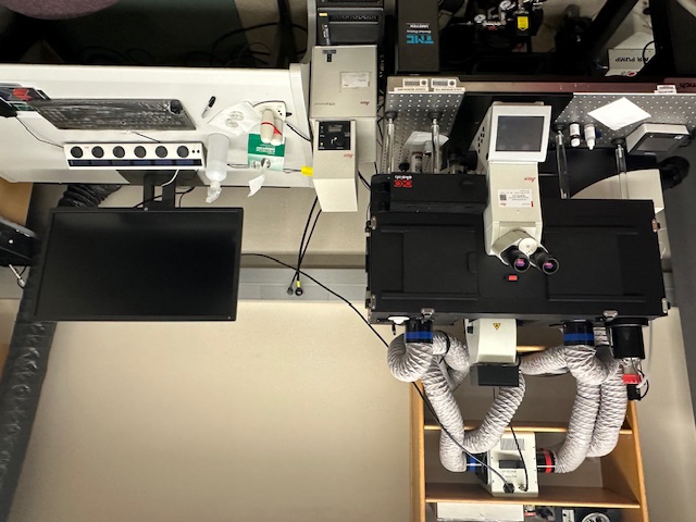
The Leica Microsystems SP8 confocal live-cell imaging system has the following features and capabilities. It is built around the Leica prism-based spectral detector system on a motorized inverted microscope stand. Instead of using emission filters, this uses a prism to disperse the emitted fluorescence, then movable mirrors to collect the desired wavelength range at the detectors. The detection range is adjustable in 1nm increments from 400-750nm. There are 4 internal detectors, two photomultiplier tube and two hybrid GaAsP-Avalanche Photodiode (HyD), which can be used simultaneously. The HyD is more sensitive and has lower noise than the PMT. There is also a transmitted light detector for simultaneously capturing a brightfield image with the fluorescence channels.
The SP8 has a Tandem Scanner which is switchable between galvo- and resonant-driven point scanning. The galvo scanner can scan a 512 by 512 field at 7 frames per second (fps). It can scan at up to 8192 by 8192 frame resolution. It also has scan zoom from 0.75 to 48x, with scan field rotation. The 8KHz resonant scanner does 28fps at 512 by 512 and has a maximum frame resolution of 2496 by 2496.
For fluorescence excitation there is a combination of an Argon gas laser and several diode lasers. These are the laser lines available: 405, 442, 458, 488, 514, 561, and 633nm. Using these lasers with the LAS X MicroLab software module provides FRAP and FRET capabilities.
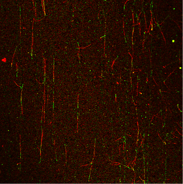
This SP8 system also has LIGHTNING super-resolution. LIGHTNING controls the acquisition parameters, analyzes the noise within the image and deconvolves the resulting scans to achieve lateral resolution down to ~120nm when using the 63x/1.4na objective. LIGHTNING also improves image quality with the other confocal scanning objectives, which include 10x and 20x.
The Digital Light Sheet (DLS) is performed after inserting a detection objective and mirror device into the transmitted light path. This system has 5, 10 and 25x detection objectives and the corresponding illumination objectives. The light sheet is produced by laser scanning onto the mirror device. The specimen is then moved through the light sheet while collecting images on a PCO Edge 5.5 sCMOS camera.
The microscope is fitted with an incubation enclosure, heated stage with CO2 control controlled via the OkoLabs incubation system for live-cell imaging.
The user interface with the confocal is a multi-knob control panel for some of the more common scanning parameters and a high power workstation running Leica LAS X. There are software modules for setting up multi-channel image acquisitions, FRAP, FRET and 3D Visualization.
Confocal
- General workflow for confocal use: WorkFlow PDF
- Operational Training Manual
- SP8 Quick Start Instructions: Quick Start
Lightsheet Mode
- Sample Preparation tips: PDF
- Leica SP8 Lightsheet Training
 |